Cell invasion into a 3D ECM requires substantial cellular traction forces as well as the degradation of ECM. We are interested in how filopodia gain traction forces from the surrounding collagen fibers in the degradable ECM. Thereby, to create the overall computational model, we integrated four modules, each capturing a different physical aspect influencing migration: 1) filopodia penetration dynamics; 2) intracellular mechanics; 3) reaction-diffusion mass transfer; and 4) structural mechanics of ECM fiber networks. We successfully compared our model with experiments of 3D HUVEC migration for diverse ECMs with different pore sizes and stiffness. Finally, our model reveals the degradation of ECM fiber network plays an important role in filopodia penetration dynamics during both tugging and contractile phases.
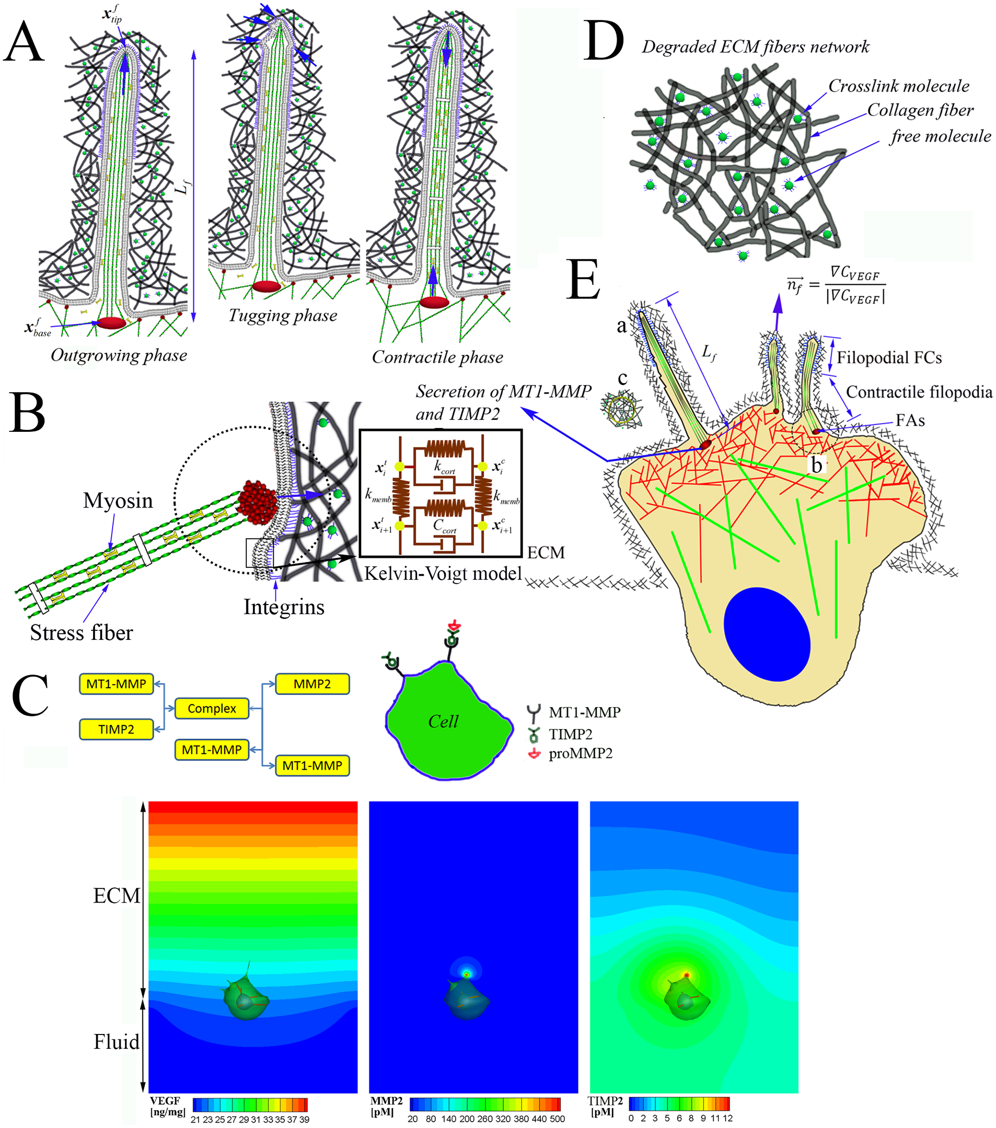
Four modules for the cell invasion dynamics. A) Filopodia dynamics showing outgrowing, tugging and contractile phases (see ‘a’ in E). B) cellular membrane mechanics showing the membrane is not only connected by actin stress fibers, but also anchored to elastic ECM fibers by forming focal adhesions (FAs) (See ‘b’ in e), and viscoelastic behaviors in cellular membrane is modeled using Kelvin-Voigt model. C) Schematic diagram of MMP-2 activation and selected simulation results of VEGF, MMP-2 and TIMP-2 concentration distributions using simplified model for MMP-2 activation. D) a magnified schematic of ‘c’ in E) showing the elastic ECM fiber networks is organized with collagen fibers, crosslinking molecules and free (or non-crosslinked) molecules. E) An integrated schematic representation of MT1-MMP and TIMP-2 secretions at the membrane near the roots of filopodia; directions of outgrowing filopodia are guided when tips of filopodia sense local gradients of VEGF toward a chemotactic cue.
Related publications
Kim, M.-C., Whisler, J., Silberberg Y.R., Kamm, R.D., and Asada, H.H. “Cell Invasion Dynamics into a Three Dimensional Extracellular Matrix Fibre Network,” PLoS Computational Biology, Vol. 11, Issue 10, e1004535, October 2015.